A Descriptive Analysis of Kinematic and Electromyographic Relationships of the Core During Forward Stepping in Beginning and Expert Dancers
Steven J. Chatfield, Ph.D., Donna H. Krasnow, M.S., Amanda Herman, and Glenna Blessing, M.F.A.
Journal of Dance Medicine & Science, Volume 11, Number 3, 2007.
Abstract
While electromyographic (EMG) and kinematic data in dance are accumulating, to date these data have raised more questions than they have answered. The purpose of this study was to introduce ensemble averaging into this body of literature as a way of dealing with the high levels of within-subject and between-subject variability that have been previously reported. This study also introduces analysis during a forward weight shift, an analysis currently absent from the literature. Three collegiate novices (18.7 ± 0.6 years of age) and three expert dancers (27.7 ± 5.5 years of age) were studied in-depth. EMG data were collected continuously at 600 Hz for analysis of onset of activity for abdominal and erector spinae muscles. Kinematic data were collected continuously at 120 Hz from markers on the acromion and the greater trochanter for analysis of the verticality of the trunk. Data were collected continuously for over 4 seconds to include: baseline data prior to movement on a right legged balance, data for movement into plié fondu on the right leg, data for a forward step to the left leg, and baseline data at resolution on a left legged balance. For analysis, data were synchronized by time using onset of vertical ground reaction forces recorded by a force plate under the initial stance leg. All participants were tested on two separate days to assess day-to-day variability. Fifteen trials were collected on each day for each individual. Ensemble averaging of continuously recorded data was used to create line graphs for visual inspection, first to compare day-to-day congruence for each individual, next to assess within group variability, and finally to compare composite graphs between groups. Day-to-day variations for each individual were minimal. Differences were seen between members of the Beginner group but not the Expert group. Between group comparisons revealed the following differences: Experts appeared to use an anterior core support strategy while Beginners appeared to use a posterior core support strategy, Experts displayed less EMG and kinematic variability than Beginners, and Experts maintained a more vertical posture throughout. Surprisingly, even though Experts were more vertical, they demonstrated the same amount of overall anterior-posterior sway as the Beginners. This finding leads to discussion of the dynamic nature of neuromuscular coordination patterns in maintenance of verticality. Issues surrounding the inability of statistically constructed models of human kinematic data to accurately represent individuals in groups are also discussed. Finally, applications of these findings to teaching and learning are offered.
The understanding and investigation of muscular activity and skeletal alignment in dancers has been a subject of interest for educators and researchers for centuries, going back to Weaver in 17211 and Blasis.[2] In the last century, more extensive descriptions of alignment and body use can be found in the literature.[3,4] However, it is only in the last 25 years that measurement tools such as electromyographic and kinematic analysis have been used in dance science research. Several studies have begun to shed light on muscle action and skeletal alignment during dance-specific movement.[5-15]
In 1992 Mouchnino and colleagues11 compared experienced modern dancers to subjects with no previous athletic or dance training. The movement task was a well-known dance movement, dégagé ŕ la seconde to 45° from turned out first position. All subjects performed 4 trials with each leg in a random sequence. In paradigm 1, the subjects were asked to do the movement as fast as possible, and maintain the resulting balance for a few seconds. In paradigm 2, they were asked additionally to keep the trunk vertical during the task. The movement was defined in two phases, the ballistic phase in which the gesture leg initiated the dégagé, and the adjustment phase in which the body found a new balance on the supporting leg. Mouchnino and colleagues[11] noted four differences. First, dancers arrived at the final balance at the end of the ballistic phase, and thus had a very short adjustment phase, non-dancers had two distinct phases, including a long adjustment phase. Second, dancers had anticipatory responses to the dégagé, whereas the non-dancers had compensatory responses, that is, in the dancers, the muscles of the supporting leg and trunk fired prior to the muscles of the gesture leg. Third, dancers minimized the center of gravity displacement to the supporting leg, whereas the non-dancers showed a larger displacement of the center of gravity. And fourth, the subjects demonstrated two distinct movement strategies. The dancers demonstrated a translation strategy, in which the hips remained level and the trunk shifted as one unit over to the new support base. The non-dancers demonstrated an inclination strategy in which the hips and head tilted to accommodate the weight shift to the new support base and which resulted in a bending of the trunk to keep the head vertical. They note that in the translation strategy, only two joints (the ankle and hip) need to be regulated, and their adjustments are equal and opposite, whereas in the inclination strategy, there are multiple joint adjustments, making the resulting action less predictable and consistent. Mouchnino and colleagues11 suggested that these differences account for much less variability in individual dancer’s trials and may be the result of long years of training.
Two more recent studies conducted electromyographic analysis of professional ballet and modern dancers, examining muscle use in stance, demiplié, and grand plié. In a 1994 study by Trepman and coworkers,[13] dancers performed five repetitions of a 6-second demi-plié, which was analyzed in two phases, the lowering phase and the rising phase. In a 1998 study by Trepman and associates,[14] dancers performed five repetitions of an 8-second grand plié, which was analyzed in four subdivided phases: early lowering phase (start to heel-off), late lowering phase (heel-off to mid-cycle), early rising phase (mid-cycle to heel-on), and late rising phase (heel-on to end). These studies found that as groups, ballet and modern dancers showed different patterns of muscle use. Furthermore, variability was seen both within each subject as well as between subjects of the same group. Interestingly, it was found that the early lowering phase of the grand plié (often thought to be the same as the lowering phase of a demi-plié) was performed differently during demi-plié and grand plié. The grand plié engaged more muscles (adductors, hamstrings, quadriceps, and tibialis anterior) in the early phase. The demi-plié was primarily quadriceps controlled. These findings yield indications that:
1. movements that appeared similar in skeletal alignment employed varying neuromuscular strategies, and
2. within any given strategy, there was regularly occurring variability across trials.
Knowing that the relationship of kinematic and electromyographic data in dance varies between individuals of similar expertise and, more importantly, even varies from trial to trial within the performances of the same individual, the intent of the current study was to employ both within and between subject analysis. In doing so, each individual’s data were preserved for analysis. This differs from study designs that rely solely on group analysis. In group analysis, individual data are sacrificed into anonymity through group averaging and other statistical procedures.
To date, all of the studies conducted have been on dancers who are either standing still or moving without traveling through space, as in stepping. The transfer of weight from one foot to the other across an open stance is a major component of dance that has not yet been analyzed using kinematics and electromyography. The purpose of this study was to investigate the relationship between select muscle recruitment and skeletal alignment (verticality) while performing dance movements involving an initial one-legged (right) balance, a forward shift of weight through space, and a resultant onelegged (left) balance at resolution. We expected to find that when compared to beginning dance students, expert dancers performing this movement would demonstrate the following: 1. maintenance of a more vertical posture, and 2. more frequent abdominal EMG responses.
Methods
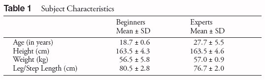
The subjects in this study were three expert dancers with mixed expertise in ballet and modern dance who were teaching and performing in a professional capacity, and three dance students with beginning level exposure to ballet and modern dance in a university dance program who were matched by height and weight with the expert dancers. Table 1 provides subject descriptions. Beginners averaged 3 hours of technique class weekly with no regularly scheduled rehearsals and no other regular physical training. Experts averaged 6 hours weekly of advanced technique class and 12 hours weekly of regularly scheduled rehearsals. Experts were not involved in any other regular physical training. Human Subjects Approval was received and all participants signed Informed Consent forms.
Testing consisted of a single step from the right foot to the left foot. Each subject’s leg was measured from the greater trochanter to the lateral malleolus and this distance was marked on the floor. This defined the distance of the forward step they were asked to perform as relative to each participant’s stature (see “leg/step length” in Table 1). The movement was demonstrated for the subjects and scripted instructions clarified that the movement was to be done while maintaining vertical alignment. In each trial, the subject did two movements in 1 second timed to an audible metronome set at 120 beats per minute. These two movements were sandwiched by baseline data at the start and baseline data at resolution. At the start, prior to moving, each subject stood ready on a one-legged balance (right leg) to establish at least 1 second of baseline data. On the first count after being cued in, the subject performed a parallel plié on the supporting right leg while reaching the gesture (left) leg forward. On the second count, the subject shifted weight forward (the distance of their trochanter to lateral malleolus measurement) onto a straight left leg, drawing the right leg to a low parallel gesture. The subject then maintained balance on the second leg to record baseline data at resolution. This created 4 phases to the test movement that will be referred to throughout this report:
Phase 1. baseline-at-start,
Phase 2. plié fondu,
Phase 3. shift of weight, and
Phase 4. baseline at resolution.
Four seconds of continuous kinematic and electromyographic data were collected beginning prior to the movement and continuing into the resolution phase. A force plate was used under the initial stance leg to record vertical ground reaction forces. For analysis, the onset of vertical ground reaction forces was used to synchronize all data by time. Baseline data at the start provided information against which phase changes associated with the movements could be seen.
For the kinematics, reflective markers were placed on the acromion of the scapula and the greater trochanter of the femur (acromio-trochanter segment). Kinematic data were collected using a 4 camera PEAK System (Peak Performance Inc., Colorado, USA). Space was calibrated to within a maximum of 2 mm error for a 1 m long wand. Threedimensional data were collected at 120 Hz. The 3-D position data were low-pass filtered between 4-8 Hz using a fourth order dual-pass Butterworth filter and recorded at 400 Hz.
Electromyographic data were collected using bipolar surface electrodes (DE-02, Delsys, MA, USA). EMG signals were on-line preamplified (x 7000), analog filtered (20-7000 Hz), and digitally converted at 600 Hz using the Associated Measurement Laboratory data acquisition system (AMLAB Inc., Sydney, Australia). For analysis, EMG signals were full-wave rectified, low-pass filtered at 6 Hz using a fourth order dualpass Butterworth filter, and recorded at 600 Hz. Abdominal electrodes were placed bilaterally in the center of a triangle formed by the inguinal ligament, a line from anterior superior iliac spine to umbilicus, and the midline.16 Erector spinae electrodes were centered over the belly of the muscle at the level of the second lumbar vertebra.
Each subject was tested on two separate days to assess day-to-day variability. Testing continued each day until data for 15 successful trials were collected. Data were collected in the motor control lab in the middle or late day. No systematic warm-up or any other preparation was given to participants. Testing sessions typically lasted approximately 90 minutes. During the initial 60 minutes, subjects were first introduced to the lab team and briefed on the testing protocol. Then the distance between their greater trochanter and lateral malleolus of their right leg was measured and footprints were manufactured and placed on the force plate and resolution stance space to reflect this distance. Next, they were outfitted with reflective markers for kinematic data collection and electrodes for EMG data collection. After outfitting was completed, the actual movement testing began and took approximately 30 minutes.
Analysis
Line graphs were created for visual inspection. These graphs contained continuously collected kinematic and EMG data starting with pre-movement baseline data and including data over the full time course of the movement as well as baseline data at resolution. All data were ensemble averaged to create one graphic representation of multiple trials. Ensemble averaging is the name given to the average patterns that can be generated for repeated trials of any continuously recorded variable. An ensemble average is generated by calculating the mean and standard deviation of a variable for every data acquisition interval for repeated trails of the same activity. For use in ensemble averaging, data need to be continuous values collected over the entire duration of the sample activity. Means and SD’s for each data acquisition interval are generated by ensemble averaging and plotted on line graphs for the entire time period of the sample activity.
For this analysis, graphs for each parameter were constructed using the ensemble average of 15 trials for each subject for each day. After visual inspection of the graphic waveforms ruled out day-to-day differences for each subject, graphs for each parameter were made that combined results for day 1 and day 2. These graphs displayed ensemble data for all 30 trials of each subject and were used to assess similarities and differences between participants within each group. Finally, ensemble graphs representing group performances were made for each parameter using all 90 trials for the beginners and all 90 trials for the experts. In all instances the ensemble averages were represented by the middle line of three lines on each graph. The ensemble average was bordered above and below by lines representing plus and minus one standard deviation.
The term “sway” was used to describe the change in degrees of verticality occurring at the intersection of a vertical line and the line formed by the acromio-trochanter segment. Graphs for sway and EMG responses were aligned for visual inspection in a time-synchronized format, using the onset of vertical ground reaction forces to establish a relative zero across the trials.
Kinematic Results and Discussion
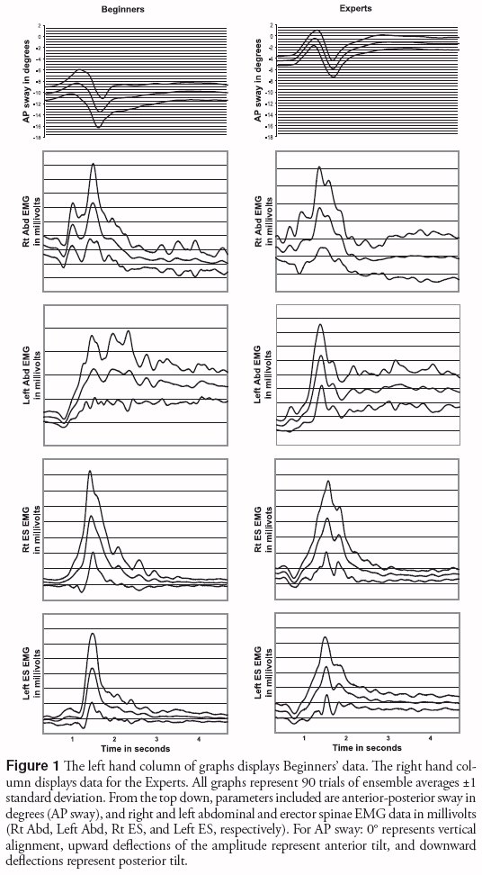
In describing sway, two factors were examined: 1. acromio-trochanter position relative to vertical at any point in time, and 2. the shape of the acromio-trochanter segment waveform over time. Figure 1 displays ensemble data representing 90 trials for each group and for each parameter. Vertical ground reaction forces were used to align movement onset at 500 msec for each parameter in Figure 1. These 500 msec of data pre-movement represent the baseline against which phasic onsets of waveforms were assessed.
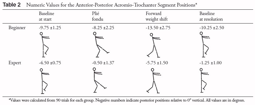
Anterior-posterior sway of the acromio-trochanter segment (AP sway) is represented in the top graph of Figure 1 for each group. Mean baseline values for the initial 500 msec of these waveforms are -9.75° ±.1.25° for Beginners and -4.50° ± 0.75° for Experts. In other words, the “ready” posture for Beginners incorporated a 9.75° posterior tilt for the acromio-trochanter segment relative to vertical. The Experts readied themselves with a 4.50° posterior tilt.
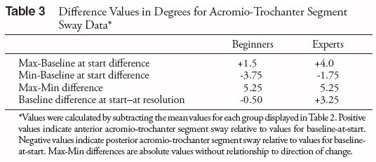
Over time, ensemble data for both Beginners and Experts produce a similar waveform for AP sway. Upward deflections of these waveforms indicate anterior sway and downward deflections indicate posterior sway (Table 2). During the plié fondu, sway is anterior. The maximum value for this anterior sway relative to the starting baseline is +1.5° for Beginners and +4.0° for Experts (Table 3). During the shift of weight, sway is posterior. The minimum, or most posterior, values relative to the starting baseline are -3.75° for Beginners and -1.75° for Experts. Another way to consider these data is that the Beginners’ sway was more than twice as large in its posterior than its anterior dimension, while the converse was true for the Experts (i.e., their anterior sway was more than twice as large as their posterior sway).
Baseline at resolution was -10.25° ± 2.50° for Beginners and -1.25° ± 1.00° for Experts. That is to say, at resolution, acromio-trochanter segments were 10.25° posterior to vertical for Beginners, and 1.25° posterior to vertical for Experts. These values at resolution indicate that Beginners ended with 0.50° more posterior tilt than they started with while Experts ended with 3.25° more anterior tilt than they started with. The absolute range of sway was calculated as the maximum minus the minimum values of AP sway. Surprisingly, this 5.25° range was the same for both groups.
In summary, a description of the Beginners’ AP sway ensemble data is that they readied themselves with a 9.75° posterior tilt from vertical, swayed 1.5° in an anterior direction during the first phase of the movement, swayed 5.25° posterior during the second phase of the movement, and resolved with a posterior tilt 10.25° behind vertical. A descriptive movement summary for the Experts is that they readied themselves with a 4.50° posterior tilt from vertical, swayed 4.0° in an anterior direction during the first phase of the movement, swayed 5.25° posterior during the second phase of the movement, and resolved with a posterior tilt 1.25° behind vertical. Compared to the Beginners, the Experts appeared to be different in that they started closer to vertical, remained more vertical throughout the movement, finished closer to vertical, and actually improved toward vertical at resolution compared to their starting position.
In the study by Mouchnino and colleagues,[11] it was suggested that dancers employ optimal strategies for shift of weight that reduce kinematic variability and compensatory activity and emphasize verticality. As described above, the current study appears to support those findings. In addition, Mouchnino and colleagues found that dancers demonstrated reduced kinematic variability during repeated performance of the same movement. The current study supports these findings. In the current study, the reduced variability for the Experts can be seen in the relative nearness of the lines representing ±1 standard deviation to the mean AP sway in Figure 1 as compared to the spread of the SD lines for the Beginners. As can be seen in Table 2, SD values for Beginners during baseline-at-start, maximum, minimum, and baseline-at-resolution range from 164% to 250% of comparable values for the Experts.
However, Beginners and Experts were not different in the overall range of AP sway that they demonstrated. Both groups swayed 5.25° as calculated by subtracting the maximum and minimum values of their AP sway characteristics. While the Experts swayed just as much as the Beginners throughout the overall time course of the movements, they did so around an axis that was closer to vertical throughout.
EMG Results and Discussion
Visual analysis of EMG findings focused on the onsets of phasic bursts. From this analysis the frequency of phasic EMG responses was assessed and select temporal characteristics of the wave forms were described (i.e., the timing of bursts relative to one another and to the time course of the movement).
Both Beginners and Experts demonstrated 100% response rates for abdominal and erector spinae EMG bursts. In other words, both Beginners and Experts displayed robust abdominal and erector spinae muscle activation responses during every trial. Examination of the Beginners’ ensemble EMG data (Fig. 1) reveals that the right abdominal EMG (R Abd EMG) ensemble response shows a double burst pattern with the first, lower amplitude spike coincident with the anterior sway during plié fondue. The second, higher amplitude R Abd EMG burst occurs later, in synchrony with the first burst of the left abdominal EMG response (L Abd EMG). These aligned R and L Abd EMG bursts occur during the final phases of the posterior sway, after toe off from the thrusting leg and during deceleration of forward momentum on the new support leg, just before the beginning of the anterior sway leading to the resolution baseline. The L Abd EMG then exhibits a pattern of multiple bursts during resolution.
For the Experts, R Abd EMG and L Abd EMG responses show fairly unified single bursts that appear to be relatively synchronous with one another and occur during the forward weight shift, beginning midway through the posterior sway. Both the R and L Abd EMG responses for the Experts then have a minor second burst on the down slope of the first that coincides with the transition into, and the initial phases of, the anterior sway that leads to movement resolution as stability is regained after the forward step is completed.
These minor second bursts for the Experts are not as pronounced as the double burst for the Beginners’ R Abd EMG or the multiple bursts spread across the resolution baseline for the Beginners’ L Abd EMG. In comparison with the Beginners, the Experts’ overall Abd EMG activity appears to be more synchronized bilaterally and more optimally sequenced in time to anticipate and transition into recovery from the posterior sway phase of the forward stepping movement.
A converse pattern of unitary versus multiple bursts exists for the groups for erector spinae EMG responses (R ES EMG and L ES EMG). For the Beginners, R and L ES EMG bursts appear synchronous and demonstrate a single focused burst during the weight shift, before their aligned R and L Abd EMG bursts, as though a posterior muscular synergy was the focus of the neuromotor strategy and antagonistic anterior responses acted to modulate the posterior response. The Beginners’ ES EMG responses preceded their Abd EMG responses by approximately 50 msec. Their ES EMG bursts occurred during the early phase of the posterior sway. Their collected Abd EMG responses occurred at the end of the weight shift as posterior sway reversed and the anterior sway leading to resolution was beginning. In other words, their collected Abd EMG bursts began during the development of the final anterior sway, which arrived at a relatively stable baseline of 10.50° posterior acromio-trochanter segment deviation from vertical during movement resolution on a one-legged balance. To the naked eye, this EMG pattern was associated with a movement that looked like a backward bowing during weight shift, followed by an impulse forward, which persisted as a jerky resolution preventing them from falling backward out of the final balance.
This response is plausibly explained as a compensatory hip strategy as described by Horak and Nashner.[17] The hip strategy is a compensatory reaction to loss of balance from posterior sway generated by a forward thrust. The ES EMG responses could be part of a synergy of posterior muscles actively backward bending the body to form a bow from head to foot that sends the body’s overall center of gravity forward to counterbalance the posterior displacement of the head and shoulders. The Abd EMG responses could be part of a synergy of anterior muscles to reduce the bowing of the body and bring the center of gravity back on top of the stance to resolve the backward bowing.
By contrast, the Experts’ ES EMG responses occurred approximately 150 msec after their Abd EMG responses. Their collected Abd EMG responses occurred in the midst of the posterior sway coinciding with the forward weight shift. Their ES EMG bursts began during the development of the anterior sway which arrived at development of a relatively stable baseline during resolution with 1.25° posterior acromio-trochanter segment deviation from vertical.
Theoretically, the Experts’ dynamic series of EMG responses seems to describe an optimal neuromotor strategy in which Abd EMG responses during the posterior sway are sequenced in time to anticipate the anterior sway that leads to resolution. This could be part of an anterior muscular synergy that helps stabilize the acromio-trochanter segment near vertical on top of a straight standing leg. The ES EMG indicates muscle activity during the anterior sway as part of a posterior muscle synergy to decelerate the forward sway of the acromiotrochanter segment to reach movement resolution and reinforce verticality of the acromio-trochanter segment on a straight standing leg with fully extended hip and knee joints.
Summary Discussion
We expected to find that when compared to beginning dance students, expert dancers performing a dance movement incorporating a forward weight shift would demonstrate: 1. maintenance of a more vertical posture and 2. more frequent abdominal EMG responses. On the first point, the Experts did demonstrate a more vertical preparation, execution, and resolution of the movement. However, surprising to us in this regard was that their overall range of sway (5.25°) was the same range of sway seen in the Beginners’ ensemble results. We found this surprising because, in retrospect, we realized that we had assumed maintaining a more vertical acromio-trochanter segment would be correlated with reduced sway overall. Apparently, this is not the case.
While this magnitude of dynamic AP sway for the experts was unexpected, it is interesting to theorize how this dynamic alignment might be a desirable strategy for dancers. First it should be noted that in real time this magnitude of AP sway is not noticeable to the naked eye. To live observers, and when viewing video footage of their performances in real time, the Experts “appeared” to maintain their verticality throughout the movement. The dynamic nature of this sway phenomenon runs counter to concepts of stiffening joints to “hold” postures during movement. Rather, a dynamic conceptualization of responsive posture during movement suggests active, ongoing kinematic and neuromuscular relationships between automatic core support mechanisms, training adaptations, and voluntary intentions.[18] As Luttgens and Hamilton state, “posture influences all we do and…it is not a static but a dynamic configuration.”19
It is interesting to note that, based on their acromio-trochanter segment sway patterns over time, the ensemble data for both Beginners and Experts in this study appear to display kinematic similarities to normal gait patterns described by Winter.[20] The ensemble results in this study describe forward tilting step initiations with backward tilting decelerations of forward thrust to arrest forward stepping momentum after weight shift and transition to a final forward tilting adjustment to establish erect posture on a straight standing leg at movement resolution. However, later in this discussion, the representativeness of the Beginners’ ensemble group model will be examined and questioned. It seems that all of the Experts do indeed display the characteristics that Winter describes as normal while 2 of the 3 Beginners do not. This could be explained by the fact that the movement used in this study was not a “normal gait” pattern. It involved stylized arm, leg, and torso use. While the Experts appear to use a “normal” gait pattern in terms of acromio-trochanter segment sway, even during this stylized movement, perhaps the novelty of the movement explains why 2 of the 3 Beginners failed to demonstrate a “normal” gait pattern for acromio-trochanter segment sway.
Further, based on the analysis of their Abd and ES EMG responses, it appears the two groups in the current study accomplish similar kinematic changes with different, contrasting neuromotor strategies. This kind of motor equivalence is a commonly accepted phenomenon within the motor control literature and has been noted in the dance literature as well.[6,12] Understanding that different individuals might accomplish similar movements through differing neuromotor strategies is important to factor into the use of generalized neuromotor coaching strategies. Many espouse an idealized, optimal approach to initiating and executing a given movement. However, there may be no such thing as an “ideal” individual to which an ideal model applies. For those who are not situated ideally, optimal performance may be elicited by very different neuromotor coaching. “It should…be understood that no single ideal postural model is appropriate for all individuals. Instead, there must be an understanding of the principles that govern efficient posture. These principles must then be applied to each individual.”[19]
The studies by Trepman and associates[13] and Trepman and coworkers[14] found variability within and between subjects during dance movements. The current study supports these findings. Because of this within subject variability, large sample pools with collapsed data analysis may not be the most informative approach to a better understanding of dancers’ strategies in complex tasks. Individual profiles are lost if only group analysis is performed. Future research in this area will most likely benefit from repeated measures designs that support ensemble averaging and mixed within and between subject analysis, so that a full depiction of the variations of individual strategies can be examined alongside group models.
For example, as mentioned earlier, analysis in the current study proceeded from individual data to group ensembles. In general, individual data from the first day closely matched data from the second day and two-day ensembles for individual data matched group ensemble data quite well. However, two features worthy of discussion were seen, and in a third instance, a remarkable difference between individual and group data was found.
The two noteworthy features of individual data as opposed to the group ensemble data seen in Figure 1 were:
1. A decreased rate of change over time as seen in the upslope of the Abd EMG burst at onset and a widening of the duration of Abd EMG spikes in ensemble versus individual data for Experts, and
2. An artificial smoothing of directional characteristics within ensemble versus individual kinematic waveforms for Beginners.
When examining individual Abd EMG bursts it is probably safe to say that, in general, each Expert had a greater rate of increase during the upslope of Abd EMG spikes at onset and each Expert had Abd EMG spikes that took a shorter period of time overall than the Experts’ ensemble group graphic represents. Minor variations between Experts operated to blunt the group ensemble rate of increase over time for the upslope of their spikes and, in addition, widened the group ensemble duration of spikes when compared to each Expert’s individual data. Even though this increased upslope and duration of the Experts’ ensemble data are blunted by the group statistic, the increased rate of upslope at onset of Abd EMG and the shortened time course of Abd EMG bursts can still be observed for Expert versus Beginner group graphics for both R and L Abd EMG data in Figure 1. In other words, even though these features are blunted for the Experts in the group model, these differences in the Experts’ ensemble group model are still fairly dramatic in contrast to the Beginner’s ensemble group model.
Seen on an individual basis, the Beginners’ Abd EMG’s were erratic, sometimes bursting unpredictably throughout the movement with no apparent coordination between R and L Abd EMGs and with repeated spikes distributed broadly through time, suggesting that their EMG responses did not have a stable relationship with major sway characteristics such as sway reversals. By contrast, the Experts’ Abd EMG responses were highly stereotypical, tightly focused, and reliably coordinated with sway characteristics of the movement.
The ensemble AP sway data in Figure 1 is represented by what appears to be a smooth, continuous line indicating that once a direction for sway had been established, it was consistent. However, when individual graphs for AP sway are examined, Beginners’ sway patterns are not as smooth as the Experts’. The Beginners have multiple “movement units” within their patterns, as well as greater deviations across trials. This concept of “movement units” has been adapted from von Hofsten and Ronnqvist[21] who define a movement unit as a directional shift in the slope of the waveform. A clear reversal of the slope was required to count as a movement unit (in other words, if the amplitude came to a plateau before re-establishing the preceding slope, it did not count as a movement unit). The Beginners in this study demonstrated multiple movement units within a given trial. By contrast, the Experts had virtually no extraneous movement units. Once the Experts established a direction of sway, their movement proceeded smoothly through that direction into a transition to either the next phase of their overall sway pattern or to movement resolution. Some of these movement unit characteristics can be seen in Figure 1 in the spread of the lines representing the standard deviations from the ensemble means. This spread is greater for the Beginners and reflects the variability generated by multiple movement units. These outcomes are in agreement with Spriggs and colleagues[22] who measured jerk (i.e., rate of change of acceleration patterns) and found that beginning, advanced, and expert dancers demonstrated increasing levels of smoothness, or decreased jerk, during performance of a dance movement. Differences in performance variability is a commonly reported finding in longitudinal studies of learning that document progressive skill acquisition and in cross-sectional studies of differences between novice and skilled performers.
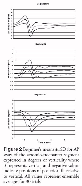
The third and probably the most significant difference between the individual and the group data is that the kinematic pattern fabricated by ensemble averaging for the Beginners does not represent the data of two of the Beginners. Dufek and associates[23] demonstrated that statistically constructed models of human kinematic data do not necessarily represent individuals in the group. To understand the motor strategies of individuals, single subject or within subject designs and analysis are required. Results of the current study provide examples of this statistical phenomenon. For example, all three of the Beginners’ AP sway graphs are displayed in Figure 2. The overall shape of the waveform through time for the top graph, for Beginner 1, is similar to the ensemble graphs for each of the Experts and for the group ensemble graph for the Beginners. The middle graph, for Beginner 2, differs from the Experts and the Beginners’ group ensemble graph in that the primary direction of the sway is anterior, without a reversal that goes posterior further than the baseline at the start. Beginner 2 tilts forward during the step and then releases that forward tilt to arrive at resolution. In other words she does not oscillate forward and backward of her starting axis like all of the Experts and Beginner 1 does and like the Beginners’ group ensemble graph indicates.
The bottom graph in Figure 2, for Beginner 3, does not have any resemblance to the anterior-posterior-anterior sway seen for the Experts, Beginner 1, and the Beginners group ensemble graph. Instead, it consists of an initial sway in the posterior direction followed by an anterior sway to resolution. Again, like with Beginner 2, there is no oscillation around her starting axis, but in this instance, the sway is entirely posterior to the starting axis, just the opposite of Beginner 2’s variation.
Being the converse of Beginner 2’s AP sway, these two data sets explain how ensemble averaging of Beginner 2 and 3 creates the anterior-posterior-anterior sway pattern seen for the Experts and Beginner 1. Beginner 2 contributes the anterior sway at the beginning of the step, and Beginner 3 contributes the posterior sway prior to movement resolution. The diversions from the group model seen in Beginner 2 and 3’s data bring up an important issue that needs to be addressed in group analysis of inherently variable phenomena, that is, individual patterns need to be assessed along side group modeling in order to fully understand the data.
In an elegant and stunning work, Dufek and coworkers[23] performed both group and single subject analysis on the same data set to assess the effects of movement experience on impact forces during jumping and running. They concluded that “The group models were not representative of any of the individual subjects’ performances and indicated that group models can describe a mythical ‘average’ performer who in fact is not representative of any of the actual performers.”[23] They recommend that researchers interested in the performance of individuals carefully evaluate experimental design before automatically using traditional group evaluation procedures. Keppel[24] clearly demonstrates how within-subjects designs control for individual variability. He goes on to say that in addition to an increase in the efficiency of data collection and analysis resulting from use of within-subject versus group designs, within-subject designs have become the designs of choice in studies of learning and transfer.
In summary, the Expert dancers in this study exhibited a smooth, dynamic, and stereotypic movement strategy that appeared to incorporate anticipatory responses during forward stepping to the balance requirement at resolution of the movement task. In contrast, the Beginners demonstrated a strategy that was jerky, had high variability from trial to trial, showed between-Beginner differences, and involved apparently compensatory responses to threats to balance during and at resolution of the forward stepping movement task. The Expert dancers were closer than the beginners to vertical alignment before, throughout, and at resolution of this movement task. Electromyographic responses suggest that the Experts optimized the work of core stability with efficient, well timed bursts of core muscle activity that helped preserve the acromio-trochanter segment’s relationship to vertical.
Surprisingly however, while the Experts’ sway oscillated around a more vertical axis than the Beginners’, the Experts had the same absolute range of sway as the Beginners. This finding matches nicely with a dynamic conceptualization of ongoing postural configurations during movement that are normal and appropriate for forward stepping.
Applications to Teaching and Learning
The results of this study suggest applications to dance pedagogy, with a focus on teaching movement through space. By examining and interpreting the strategies of the expert dancers, we may be able to enhance the process of achieving reliably coordinated, smooth, dynamic torso movement off the standing base, with core support, during shift of weight in traveling work. The results of this investigation point out the inherent sway and ongoing dynamic adjustments to posture during forward stepping. This adaptable concept of dynamic stability stands in high contrast to neuromotor patterns that stiffen against torso sway in maintenance of vertical posture. If the smooth, dynamically responsive and reliable performance model displayed by the expert dancers in this study is the desired model, concepts such as holding or tightening to maintain vertical, and approaches that encourage restricting adaptive sway of the torso, may be counterproductive to the coordination patterns seen in this study and to optimal weight shift strategies.
How might dance educators assist dancers in developing the coordination of smooth, dynamic torso movement during shift of weight in traveling work? In an exercise designed to encourage the strategy seen in this study, a teacher could assist in the following way. During the pre-movement “ready” phase, prior to initiation, take a moment to help the dancers visualize the action they are about to perform, talk them through it using vivid detail that includes the ongoing changes needed as the demands of the movement progress. Then, with these intentions in mind, perform the action. By promoting this pre-movement anticipation and intentionality, the teacher can guide the dancer toward the discovery and use of neuromotor strategies like those observed in the expert dancers in this study.
Finally, teachers can recognize the need to encourage and support individualized work. We need to define and describe the goal of the action and then allow each dancer to explore and experiment with multiple strategies until the most effective one for that individual is realized. Attention to the task, using visualization and awareness of initiation mechanisms, may allow the system to self-organize, resulting in a reduction of unnecessary tension and increased efficiency in muscle use. For decades the practitioners of somatic practices, such as Feldenkrais and Alexander, have been approaching neuromotor re-education from this perspective of awareness, selfdiscovery, and ease without restrictive holding to achieve movement goals. The expert dancers in this study achieved the smooth, coordinated shift of weight with a motor strategy allowing sway and ongoing dynamic adjustments. Dance educators might be more effective in moving dancers toward more appropriate and efficient strategies such as those displayed by the expert dancers in this study by encouraging introspective exploration of movement, similar to the way guided experiences are structured in somatic work, and allowing each dancer’s neuromotor system to find increasingly more fluid and elegant patterns of coordination as options to their old patterns.
References
2. Blasis C: An Elementary Treatise upon the Theory and Practice of the Art of Dancing. Translated by M. S. Evans. New York: Kamin Dance Publishers, 1953.
3. Kirstein L, Stuart M: The Classic Ballet: Basic Technique and Terminology. New York: Alfred A. Knopf, 1976.
4. Lawson J: Classic Ballet: Common Faults in Young Dancers and Their Training. New York: Theatre Arts Books, 1973.
5. Barnes MA, Krasnow D, Tupling SJ, Thomas M: Knee rotation in classical dancers during the grand plié. Med Probl Perform Art. 2000;15:140-7.
6. Chatfield SJ, Barr S, Sveistrup H, Woollacott MH: Electromyographic and kinematic analysis of movement repatterning in dance. Impulse, the International Journal of Dance Science, Medicine, and Education. 1996;4(3):220-34.
7. Gamboian N, Chatfield SJ, Woollacott MH, Klug GA: Effect of dance technique training and somatic training on pelvic tilt and lumbar lordosis alignment during quiet stance and dynamic dance movement. J Dance Med Sci. 1999;3(1):5-14.
8. Krasnow DH, Chatfield SJ, Barr S, et al: Imagery and conditioning practices for dancers. Dance Research Journal. 1997;29(1):43-64.
9. Meglin J, Woollacott M: The neural choreography underlying a pirouettearabesque. Kinesiology and Medicine for Dance. 1992;14:95-105.
10. Monasterio RA: Postural adjustment for voluntary leg movement in dancers. Unpublished master’s thesis, University of Oregon, 1994.
11. Mouchnino L, Aurenty R, Massion J, Pedotti A: Coordination between equilibrium and head-trunk orientation during leg movement: a new strategy built up by training. J Neurophysiol. 1992;67(6):1587-98.
12. Ryman R, Ranney D: A preliminary investigation of two variations of the grand battement devant. Dance Research Journal. 1978/79;11:2-11.
13. Trepman E, Gellman RE, Micheli LJ, De Luca CJ: Electromyographic analysis of grand-plié in ballet and modern dancers. Med Sci Sports Exerc. 1998;30(12):1708-20.
14. Trepman E, Gellman RE, Solomon R, et al: Electromyographic analysis of standing posture and demi-plié in ballet and modern dancers. Med Sci Sports Exerc.1994;26(6):771-82.
15. Wilmerding V, Heyward V, King M, et al: Electromyographical comparison of the developpe devant at barre and centre. Presented at the 9th Annual Meeting of the International Association for Dance Medicine & Science, Tring, England, 1999.
16. Snijders CJ, Ribbers MTLM, de Bakker HV, et al: EMG recordings of abdominal and back muscles in various standing postures: Validation of a biomechanical model on sacroiliac joint stability. J Electromyogr Kinesiol. 1998;8:205-14.
17. Horak F, Nashner L: Central programming of postural movements: adaptation to altered support-surface configurations. J Neurohysiol. 1986;55:1369-81.
18. Krasnow DH, Monasterio RA, Chatfield SJ: Emerging concepts of posture and alignment. Med Probl Perform Art. 2001;16:8–16.
19. Lutgens K, Hamilton N: The standing posture. In: Kinesiology: Scientific Basis of Human Motion (9th ed). Dubuque, IA: Brown & Benchmark. 1997, pp. 445-459.
20. Winter DA: The Biomechanics and Motor Control of Human Gait: Normal, Elderly, and Pathological (2nd ed). Waterloo, Ontario, Canada: University of Waterloo Press, 1991.
21. Von Hofsten C, Ronnqvist L: The structuring of neonatal arm movements. Child Dev. 1993;64:1046-57.
22. Spriggs J, Bronner S, Brownstein B, Ojofeitimi S: Smoothness during a multi-joint movement: 2D and 3D analysis between groups of differing skill levels. Presented at the 12th Annual Meeting of the International Association for Dance Medicine & Science, New York, USA, 2002.
23. Dufek JS, Bates BT, Stergiou N, James CR: Interactive effects between group and single-subject response patterns. Hum Mov Sci. 1995;14:301-23.
24. Keppel G: Design and Analysis: A Researcher’s Handbook (3rd ed). Englewood Cliffs, NJ: Prentice Hall, 1991.