Knee Rotation in Classical Dancers during the Grand Plié
Margaret A. Barnes, M.A., T.Dip. (RAD), Donna Krasnow, M.S., Susan J. Tupling, Ph.D., and Martin Thomas, Ph.D.
Medical Problems of Performing Artists, December 2000.
Abstract
For the past two decades, dance researchers and educators have been concerned about the use of the grand plié and its potential impact on injury incidence. The purpose of this study was to investigate external longitudinal rotation (ELR) at the knee during grand pliés performed in three classical ballet leg positions, 2nd, 3rd, and 4th, by professional female ballet dancers. Light reflective markers were placed on seven lower body segments of ten volunteer subjects. Subjects performed three trials of the grand plié and one demi-plié in each of the three leg positions while being videotaped. Marker locations were digitized. Five “instants” were identified in the plié: begin, demi-plié lowering phase, grand plié, demi-plié rising phase, and finish. The ELRs at the knee joints for each instant were analyzed using an analysis of variance (ANOVA) and custom matrix analysis. Results indicate that ELR values are highest at the bottom of the movement in all positions, and that 3rd and 4th position grand pliés present higher overall ELRs than 2nd position. Because ELR at the knee has been named as one of the more serious predisposing factors to knee injury, the results of this study indicate that further investigation about the grand plié is needed, and educators should include grand pliés in the training process with caution.
Too often a dancer’s career is shortened by an injury, and often this injury is at the knee joint. [1–3] In particular, longitudinal rotations at the knee joint are thought to stretch or damage connective and contractile tissue in the knee. This damage may include: stretching and/or irritating the anterior cruciate ligaments, increasing rotary instability of the joint and knee hyperextension; stretching and/or irritating medial collateral ligaments, compromising the lateral stability of the joint; maltracking of the patella in the trochlear groove, resulting in grinding of the articular cartilage of the patellofemoral joint; tearing or rupturing of the menisci; increasing the incidence of patellar tendonitis and/or chondromalacia patella—“jumper’s knee”; and predisposing the joint to patellar dislocation.4–8 The grand plié is a dance exercise and performance skill that has been cited as being potentially harmful to the knee joint due to the associated compressive forces, excessive range of motion, and longitudinal rotations that occur at the joint during this movement. [9–11]
Several authors have argued against using the grand plié as a training exercise because of its high risk-to-benefit ratio, [2,7,9,10,12–14] and frequent pliés have been implicated as a contributing factor to chronic lower limb injuries. [14] However, grand pliés occur in some of the contemporary and classical performance repertoires. Because physical training requires both repetition and specificity of movement to ensure appropriate neuromuscular patterns, [13,15,16] it would seem unwise to eliminate grand pliés completely from the class structure if the same movements are to remain in the performance setting.
Both acute and overuse injuries incurred by ballet dancers are most often caused by physical compensations made in order to overcome anatomical limitations in hip joint ranges of motion (ROMs). [4] However, because of the standard aesthetic demands of classical ballet, dancers often sacrifice good alignment for the appearance of greater “turnout” or external rotation through the leg. [17] Combining the extreme amount of external rotation demanded by ballet technique with the nature of the plié movement itself seems to be problematic in the interest of maintaining good alignment and avoiding injury. [9,12,13] Poor alignment includes pronation of the foot, excessive anterior pelvic tilt, and excessive external longitudinal rotation (ELR) of the lower leg.4 In addition, foot pronation and poor alignment of the torso during pliés are often the causes of increased ELR. [13] However, the relationship between degree of knee flexion, ELR values at the knee joint, and leg position during grand pliés has not yet been established.
Until recently, there have been few adequate measurement tools available for detailed research on the plié. Electromyography, isokinetic dynamometry, goniometric measurements, manual techniques, and two-dimensional (2D) cinematography are some of the techniques researchers have employed to investigate how pliés are performed.
While muscular recruitment patterns, vertical torso alignment, and 2D joint ROMs have been investigated during static and dynamic demi (1st position) and grand pliés (1st and 2nd positions), [7,8,11,12,18–20] a complete three-dimensional (3D) description or mapping of the actual movements of the lower limb segments and joints from dynamic grand pliés or in crossed positions of the legs has not been made. Even classical ballet references [21] only define pliés as the bending of the knees with the legs in external rotation. Krasnow et al. suggest that further study investigating dancers in motion would be more relevant than study of dancers in static positions. [19] Considering the number of grand pliés often performed in a traditional ballet class, [10] it is rather alarming that more is not known about the movement.
PURPOSE
It was the aim of this study to collect kinematic data regarding longitudinal rotations in the knee joints of professional female ballet dancers performing grand pliés. From this information about knee joint rotations, our knowledge about knee health and predisposing factors to chronic knee injuries in dancers would be enhanced, with the hope of carrying dance teaching practices to a new level of efficiency and responsibility in the interest of career longevity and injury prevention.
The following definitions may be helpful to the reader:
• Turnout: a term used interchangeably with “external rotation.” According to The Dictionary of Classical Ballet Terminology, turnout comes from the hip, ideally with 90° coming from each of the left and right sides. [21]
• The Plié: a knee bend. In classical ballet it is usually performed with the legs externally rotated, and the torso vertically aligned and centered over both legs. The grand plié has been defined previously as a movement “in which the upright torso, spine, and pelvis are stabilized as they are lowered with coordinated hip and knee flexion, and then raised back to the starting position with hip and knee extension.” [11]
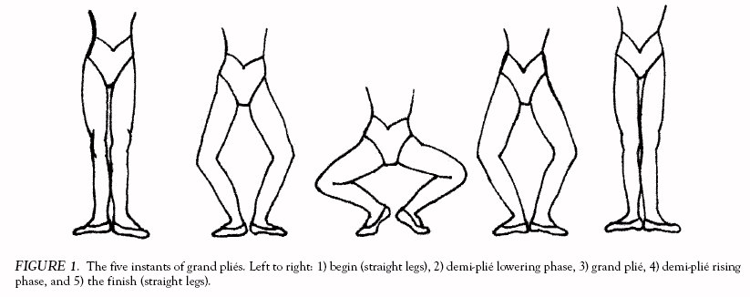
• Instant: a specific videotaped frame identified through visual inspection of the entire trial to represent one of the five moments during the grand plié to be statistically analyzed: 1) begin (straight legs), 2) demi-plié lowering phase, 3) grand plié, 4) demi-plié rising phase, and 5) the finish (straight legs).22 Figure 1 illustrates the five instants of the grand plié for the 1st position.
• The Demi-Plié Instant (DPI): The demi-plié movement is a bending of the knees with the legs turned out so that the knees are directed over the toes, and the heels remain on the floor. [21] For the purposes of this study, the DPI in the 3rd and 4th positions of the legs is defined as the body position at the last moment of contact of the heels with the floor (instants 2 and 4). Therefore, in the 3rd and 4th positions, the DPI is defined as the moment just before the front heel leaves the floor during descent and the moment just after the front heel regains contact with the floor during ascent.22 This instant was determined by visual inspection of the abduction/adduction (x-axis rotation) data for the right (front) foot. The front heel was chosen to define the DPI because it was easier to observe the movements of this heel. In the 2nd position, the heels never lose contact with the floor during the grand plié. Consequently, the DPI in the 2nd body position is defined as the position achieved at half-way between instant 1 and the GPI during descent, and between the GPI and instant 5 during ascent, measured by frame number. [22]
• The Grand Plié Instant (GPI): the moment at the bottom of the grand plié movement when the dancer changes the direction of her plié from descent to ascent.22 The GPI (instant 3) was identified by visual inspection of the data for knee flexion/extension angles of the front (right) knee. The front knee was used to define the GPI so that the same leg was used to define both the DPI and the GPI. In the case of a curve exhibiting a plateau at maximum knee flexion, the midpoint of the plateau defined the GPI.
Five research hypotheses were posed:
1. External rotation in the knee will be greatest at the bottom of the grand plié movement (GPI) when compared with both demi-plié (DPI) and straight leg stance (lowering or rising phase), regardless of plié position.
2. Of the five instants of the grand plié movement, ELR values will be greatest at the 4th position GPI (instant 3) compared with any other plié position and any other instant.
3. At any specific instant of plié trials performed in the three plié positions, the magnitude of ELR values will be greatest in the 4th position, and least in the 2nd position.
4. There will be no symmetry between the ELR values for the left and right knees of classical ballet dancers at any instant of grand pliés performed in any plié position.
5. There will be no symmetry between the ELR values in the knees of classical ballet dancers at similar instants across the eccentric (lowering) and concentric (rising) phases of grand pliés in any plié position.
METHOD
Fourteen professional female ballet dancers (mean age = 23 years, SD = 4.721 years, range = 17 years to 34 years; mean height = 167.343 cm, SD = 5.011 cm) consented to take part in the study. Following digitization and reduction, ten complete data sets were obtained. At the time of filming, all of the subjects were under contract as apprentices, corps members, soloists, or principal dancers with a large Toronto-based classical ballet company. Prior to acceptance into the sample group, all dancers completed a medical questionnaire. None of the subjects were taking time off from performing due to injury at the time of filming. Prior to filming, a series of supplementary measures were made on each dancer to record maximum passive and active external and internal hip rotations, and active knee hyperextension. Subjects also completed posttest questionnaires designed to gain information about their dance backgrounds, their histories of knee pain, and any formal training they had received in teaching ballet.
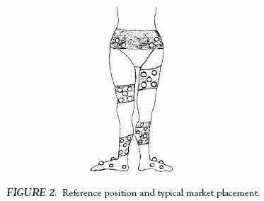
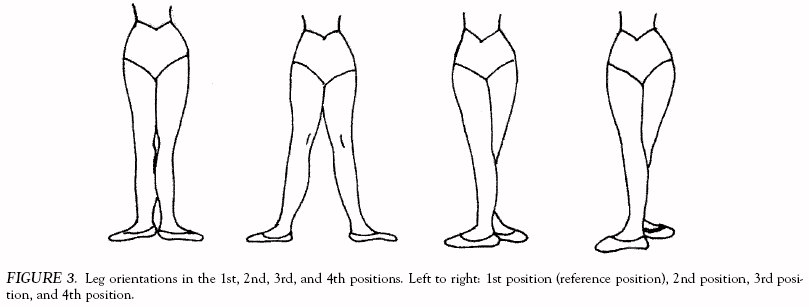
In this study body segments were modeled as rigid bodies whose 3D spatial location could be described using three sequential rotations from some arbitrary initial position. Classical 1st position (Figs. 2 and 3) was used as the initial reference position, which ensured that the video cameras were able to capture all of the reflective markers. The sequential rotations followed common anatomical human movement patterns, namely, flexion or extension first, followed by abduction or adduction, and finally internal or external rotation (also known as a body-fixed 3-1-2 Cardan angle system).23 A body-fixed coordinate system moves with the body segment and is not fixed within the filming space. The 3D location of a minimum of three points on the rigid body must be known before location of the rigid body can be found using this approach. To analyze the plié, a seven-segment model of the lower human body (pelvis, right/left thigh, right/left lower leg, and right/left rear foot) was defined. Four to five silvercolored light-reflective markers were placed on each segment (32 total) (Fig. 2).
Subject Protocol and Data Collection
All the subjects performed a self-directed 10-minute warmup prior to marker placement. During filming, each dancer performed three grand pliés and one demi-plié in each of three leg positions (2nd, 3rd, and 4th), and was filmed standing in the reference position. Figure 3 shows the orientation of the legs in the 2nd, 3rd, and 4th positions, in addition to the reference position (1st). A metronome was used to standardize the length of all trials. Plié movements were captured on two professional-quality VHS video cameras set at 5 m to the center of the filming area with a separation angle of 40 degrees. The 35-mm camera flash that signaled the dancers to begin each plié was also used to synchronize the video cameras during data reduction. An aluminum cube with 180.0-cm sides, provided by Ariel Dynamics (Trabuco Canyon, CA), was used to calibrate the filming space. Frames of the cube were recorded before and after filming each subject as a precaution against the effect of moving a camera during filming.
To maintain relative distance between markers on a given segment and the location of the markers on the segment after recording the reference position, 4-inch-wide Velcro strips were attached across the grain of two-way stretch elastic straps, and the light-reflective markers were attached to the straps using the Velcro strips. The straps were custom-fit to each dancer to accommodate differences in muscle definition and placement. In spite of these precautions, one of the subjects was dropped from the study. For this subject, thigh and lower leg rotations were so exaggerated at several moments during plié trials as to leave only two markers in view, making it impossible to obtain 3D data.
Data Reduction and Analysis
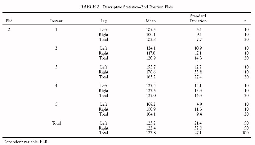
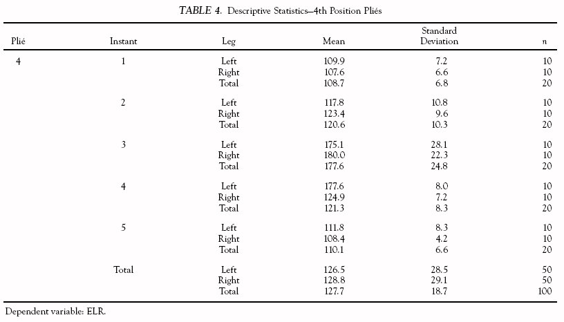
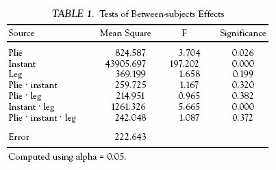
One randomly chosen trial for each subject and each foot position was selected for analysis. Marker locations for these trials, the reference position for each dancer, and the calibration cube were digitized using Ariel Dynamics software (The Ariel Performance Analysis System—APAS), which utilized both manual and automatic digitization. For plié trials, every fourth frame was captured for digitization. At a normal camera speed of 60 frames/sec, the effective digitization frame rate was 15 frames/sec. The 3D marker locations were calculated using a direct linear transformation algorithm (Ariel Dynamics). The 3D segment (rigid body) angular orientations and the corresponding joint angles were determined using specially written software using the Cardan angles that have already been discussed. Data from three subjects were dropped from the study at this time, because mathematical instabilities that occurred when the thigh abduction angles reached 90 degrees yielded data that were not in line with physiological possibility. From the ten complete data sets, the y-axis rotations for the five defined instants of grand pliés were extracted from each trial selected for analysis. The ELRs at both knee joints at each instant during the movement were analyzed using an analysis of variance (ANOVA) least squares difference (LSD) post-hoc tests, and custom contrast matrix analyses. The ELR values reported in the following plots have undergone transformations needed for the statistical analysis: to make all values positive, left knee rotations were multiplied by –1, and 100 degrees were added to both left and right knee rotations. The transformed ELR values are denoted as “ELR valuesC.” Results of statistical tests, standard deviations, and all ELR valuesC are reported in Tables 1–4.
RESULTS AND DISCUSSION
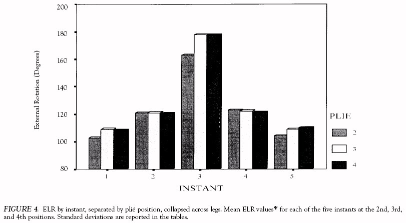
An analysis (ANOVA) of the ELR valuesC data revealed a significant main effect for instant [F(4,270) = 197.202, p = 0.001] (hypothesis 1), and positive results of an LSD analysis performed on the data indicated significant differences in ELR during straight leg stance, at DPI, and at the bottom of the movement (p = 0.05) (Fig. 4). Inspection of Figure 4 shows higher ELR valuesC associated with the bottom of the movement, regardless of leg position. The slightly higher values observed here in the 3rd and 4th position trials failed to reach significance.
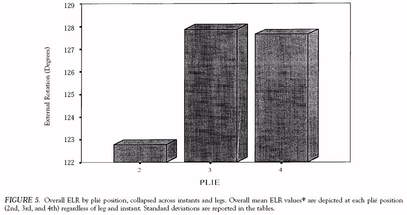
Figure 5 depicts a significant main effect for plié position [F(4,270) = 3.704, p = 0.026] (hypothesis 3), indicating that 3rd and 4th position grand pliés involved greater overall ELR valuesC than 2nd position grand pliés.
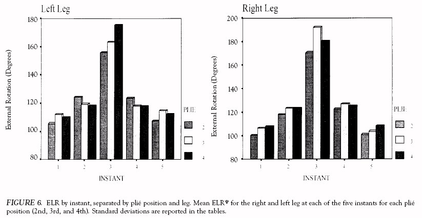
There was also a significant combined effect for instant and leg [F(1,270) = 5.665, p = 0.001] (hypothesis 4) revealed by a custom matrix analysis of the ANOVA testing. A significant difference in ELR valuesC between legs was found only at the bottom of the grand plié movement, and not at any other instant, partially supporting hypothesis 4 that there will be no symmetry between ELR rotation in the knees at any instant during grand pliés (regardless of plié position) (Fig. 6).
Statistical analyses of the data obtained in the present study found that ELR is highest at the bottom of the grand plié movement compared with any other instant, regardless of plié position, and that the ELR recorded at DPI is higher than during straight leg stance, indicating that ELR increases with knee flexion. Increased ELR has been discussed as being one of the leading predisposing factors to soft-tissue injury at the knee in dancers. Therefore, grand pliés may be more dangerous to the structure of the knee joint than demi-pliés.
The three plié positions tested (2nd, 3rd, and 4th) did not differ enough from each other with respect to the amount of ELR created at any specific instant to reach statistical significance. It is possible that the anterior pelvic tilt and posterior translation of the pelvis that was noted during some 2nd position trials added to the mean ELR at instant 3 in this position, thereby bringing the mean ELR values for all of the positions at this moment closer together. Also, because of the combined effect of patellar compression forces, which increase with knee flexion, and the high amount of ELR recorded at the bottom of 3rd and 4th position trials, grand pliés performed in these crossed positions of the legs may be more problematic than 2nd position grand pliés in compromising the integrity of the knee joint. However, although the three plié positions tested did not differ significantly from each other at any particular instant, higher overall mean ELR values (across the entire movement) were recorded in 3rd and 4th position grand pliés than in 2nd position grand pliés. Therefore, the data suggest that grand pliés in the 3rd and 4th positions present higher risk to the knee overall than 2nd position grand pliés.
At GPI, the ELR values between knees differed enough to reach significance. These values were not different enough at any other instant to reach significance. These, and the patterns observed for the thigh and lower leg segments of pliés performed in the 2nd, 3rd, and 4th positions, suggest that the grand plié is not just a continuation of the demi-plié, and that the legs experience different patterns of movement during pliés performed in different positions. This supports a similar conclusion by Trepman et al.20 that the grand plié is not an extension of the demi-plié regarding muscle use. The ELR values measured throughout were essentially symmetrical during the descending and ascending halves of plié movements in any plié position (negating hypothesis 5).
CONCLUSION
This was the first study undertaken to examine the ELR at the knee joints in situ of ballet dancers performing grand pliés in various plié positions. Furthermore, the data were obtained using 3D technology and analyses. In the process of completing the original study, a model describing lower body segment rotations during 2nd, 3rd, and 4th position grand pliés was created and the 3D segmental motions of each segment and the knee joint angle were completely described. [22]
Further study is required to overcome some of the technical details and computational challenges presented by doing rigid body studies. It would be beneficial to optimize the data collection variables for movements performed in dance (number and placement of cameras, marker diameter, direction of light sources, etc.). Fine-tuned reduction techniques that calculate present-frame Cardan angles from previousframe values while calculating a “best fit,” and that prevent the mathematical instabilities encountered in the present study when the thigh segments reached 90 degrees of abduction, would be ideal. This mathematical improvement would allow the utilization of the present system in similar applications studying the 3D movements of dancers, which would yield easily interpreted results that closely represent human movement, making the results accessible to a wide audience.
The results of this study indicate that high ELR values occur at the bottom of the plié movement when compared with other points during the movement. Other factors, such as the ratio of leg length and leg separation distance in open positions (2nd and 4th), amount of total external rotation through the leg at the start of the movement, and vertical alignment through the torso may also affect these values and would be worth studying. Further study employing 3D analysis of grand pliés in various positions is needed to determine the relationships between amount of total hip joint turnout and the ratio between leg length and width of open positions on the ELR experienced at the knee.
The findings of this study may be useful in the prevention of knee injuries in dancers. The high ELR values recorded at the bottom of grand pliés indicate that excessive repetitions of the movement may compromise knee joint stability. Also, investigating the effect of repeated use of this movement on young dancers entering growth spurts would be prudent in the interest of injury prevention in upcoming generations of dancers. The predisposing factors that are caused by ELR at the knee joint when performing grand pliés can compromise knee joint stability and are compounded over time, and muscular fatigue may increase the risk of sudden meniscus injury when the knee is in a position of extreme flexion. Therefore, caution when performing repeated grand pliés is recommended, especially in crossed positions of the legs such as 3rd and 4th. Evidently, further research is needed to examine the relationship between increased ELR at the knee and the high incidence of knee injuries in dancers.
REFERENCES
1. Chmelar RD, Shultz BB, Ruhling RO, Fitt S, Johnson MB: Isokinetic characteristics of the knee in female, professional and university, ballet and modern dancers. J Orthop Sports Phys Ther 9:410–418, 1988.
2. Gelabert R: Preventing dancer’s injuries. Physician Sportsmed 8(4): 69–76, 1980.
3. Ryman R: Training the dancer XI: The knee—Achilles heel of the body. Dance Can Magazine Spring:12–17, 1980.
4. Hamilton WG: Ballet and your body: An orthopedist’s view. Dancemagazine 6:84–85, 1978.
6. Malcolm R: The screw home mechanism and its implications for dancers. Impulse 4:253–257, 1996.
7. Wislow J, Yoder E: Patellofemoral pain in female ballet dancers: Correlation with iliotibial band tightness and tibial external rotation. Sportsmed Physiother 22(1):18–21, 1995.
8. Zarins B, Rowe CR, Harris B, Watkins MP: Rotational motion of the knee. Sports Med 11:152–156, 1983.
9. Hamilton WG: Ballet and your body: An orthopedist’s view. Dancemagazine 5:98–99, 1978.
10. Myers M: Is the grand plié obsolete? Dancemagazine 6:78–80, 1982.
11. Trepman E, Gellman RE, Micheli LJ, DeLuca CJ: Electromyographic analysis of grand-plié in ballet and modern dancers. Med Sci Sports Exerc 30:1708–1720, 1998.
12. Clippinger-Robertson K, Hutton RS, Miller D, Nichols R: Mechanical and anatomical factors relating to the incidence and etiology of patellofemoral pain in dancers. Kinesiol Dance 7(3):7–9, 1985.
13. Hecox B: Reader’s forum [letter to the editor]. Dancemagazine 11:48, 1982.
14. Milan KR: Injury in ballet: A review of relevant topics for the physical therapist. Orthop Sports Phys Ther 19(2):121–129, 1994.
15. Krasnow DH, Chatfield SJ: Dance science and the dance technique class. Impulse 4:162–172, 1996.
16. Liederbach M: Movement and function in dance. In Brownstein B, Bronner S (eds.): Evaluation, Treatment and Outcomes: Functional Movement in Orthopaedic and Sports Physical Therapy. New York, Churchill-Livingstone, 1997, pp. 253–310.
17. Weiker GG: Dance injuries: The knee, ankle, and foot. In Clarkson P, Skrinar M (eds.): The Science of Dance Training. Champaign, IL, Human Kinetics Books, 1988, pp. 147–192.
18. Clippinger-Robertson K: Principles of dance training. In Clarkson P, Skrinar M (eds.): The Science of Dance Training. Champaign, IL, Human Kinetics Books, 1988, pp. 45–90.
19. Krasnow DH, Chatfield SJ, Barr S, Jensen JL, Dufek JS: Imagery and conditioning practices for dancers. Dance Res J 29(1):43–64, 1997.
20. Trepman E, Gellman RE, Solomon R, Murthy KR, Micheli LJ, DeLuca CJ: Electromyographic analysis of standing posture and demi-plié in ballet and modern dancers. Med Sci Sports Exerc 26:771–782, 1994.
21. Ryman R: The Dictionary of Classical Ballet Terminology. Royal Academy of Dancing, London, England, in cooperation with the University of Waterloo, Waterloo, Canada, 1995.
22. Barnes MA, Krasnow DH, Tupling SJ, Thomas M: Knee rotation in classical dancers: A three dimensional videographic analysis. Master’s thesis, York University, 1999.
23. Tupling S, Pierrynowski MR: Use of Cardan angles to locate rigid bodies in three-dimensional space. Med Biol Engineering Comput 25: 527–532, 1987.
SUGGESTED READING24. Barrack RL, Skinner HB, Brunet ME, Cook SD: Joint laxity and proprioception in the knee. Physician Sportsmed 11:130–135, 1983.
25. Basmajian JV: Primary Anatomy (7th ed). Baltimore, MD, Williams & Wilkins, 1976.
26. Bergfield JA: Medical problems in ballet: A round table. Physician Sportsmed 10:98–112, 1982.
27. Clarkson P, Skrinar M. The Science of Dance Training. Champaign, IL, Human Kinetics Books, 1988.
28. Fitt SS: Dance Kinesiology. New York, Schirmer Books, 1988.
29. Klein P, De Haven J: Accuracy of three-dimensional linear and angular estimates obtained with the Ariel Performance Analysis System. Published report from the American Academy of Physical Medicine and Rehabilitation, 76:183–189, 1995 [on line]. Available: http://www. arielnet.com/newlook/adw-27.html.
30. Spiegelman J, Woo S: A rigid body method for finding centers of rotation and angular displacements of planar joint motion. Biomech 20: 715–721, 1987.
31. Swiegard LE: Human Movement Potential: Its Ideokinetic Facilitation. Lanham, MD, University Press of America, 1974.